Beranda
/ Arteries Diagram - Diagram Of Veins And Arteries In Body Hd Png Download Kindpng - The aorta is the main systemic artery and the largest artery of the body.
Arteries Diagram - Diagram Of Veins And Arteries In Body Hd Png Download Kindpng - The aorta is the main systemic artery and the largest artery of the body.
Insurance Gas/Electricity Loans Mortgage Attorney Lawyer Donate Conference Call Degree Credit Treatment Software Classes Recovery Trading Rehab Hosting Transfer Cord Blood Claim compensation mesothelioma mesothelioma attorney Houston car accident lawyer moreno valley can you sue a doctor for wrong diagnosis doctorate in security top online doctoral programs in business educational leadership doctoral programs online car accident doctor atlanta car accident doctor atlanta accident attorney rancho Cucamonga truck accident attorney san Antonio ONLINE BUSINESS DEGREE PROGRAMS ACCREDITED online accredited psychology degree masters degree in human resources online public administration masters degree online bitcoin merchant account bitcoin merchant services compare car insurance auto insurance troy mi seo explanation digital marketing degree floridaseo company fitness showrooms stamfordct how to work more efficiently seowordpress tips meaning of seo what is an seo what does an seo do what seo stands for best seotips google seo advice seo steps, The secure cloud-based platform for smart service delivery. Safelink is used by legal, professional and financial services to protect sensitive information, accelerate business processes and increase productivity. Use Safelink to collaborate securely with clients, colleagues and external parties. Safelink has a menu of workspace types with advanced features for dispute resolution, running deals and customised client portal creation. All data is encrypted (at rest and in transit and you retain your own encryption keys. Our titan security framework ensures your data is secure and you even have the option to choose your own data location from Channel Islands, London (UK), Dublin (EU), Australia.
Arteries Diagram - Diagram Of Veins And Arteries In Body Hd Png Download Kindpng - The aorta is the main systemic artery and the largest artery of the body.. We hope this picture human body artery diagram in detail can help you study and research. Learn vocabulary, terms, and more with flashcards, games, and other study tools. A wire is moved through an artery in the leg up to the carotid artery, and a small wire tube, or stent is expanded inside a narrowing of the carotid artery. Heart anatomy coronary arteries diagram. The veins also lack the elastic internal lamina that lies.
For more anatomy content please follow us and visit our website: Blood is transported in arteries, veins and capillaries. Constricted arteries oppose blood flow, and more pressure is required to push blood. The subclavian artery runs into the axillary region where it becomes known as the axillary artery. Coronary circulation is the circulation of blood in the blood vessels that supply the heart muscle (myocardium).
Arteries And Veins Of The Leg Educational Diagram Download Free Vectors Clipart Graphics Vector Art from static.vecteezy.com Learn the differences between an artery and a vein. Arteries and arterioles carry oxygenated blood _____ from the heart to the body. Blood is pumped from the heart in the arteries. This is called peripheral artery disease (pad). Anatomynote.com found human body artery diagram in detail from plenty of anatomical pictures on the internet. Diagram of the human circulatory system (infographic). There are two paired arteries which are responsible for the blood supply to the brain; Start studying review of arteries.
Blood is transported in arteries, veins and capillaries.
The tunica medica, which is the very muscular middle layer in arteries, is thinner and less muscular in veins. Circle of willis is indeed a hot neuroanatomy topic! Arteries and arterioles carry oxygenated blood _____ from the heart to the body. An artery (plural arteries) (from greek ἀρτηρία (artēria) 'windpipe, artery') is a blood vessel that takes blood away from the heart to one or more parts of the body (tissues, lungs, brain etc.). There is a point at which the anterior and posterior arterial circuits of the brain unite or anastomose. It originates from the heart and branches out into smaller arteries which supply blood to the head region (brachiocephalic artery), the heart itself (coronary arteries), and the lower regions of the body. These vessels are channels that distribute blood to the body. Anatomynote.com found human body artery diagram in detail from plenty of anatomical pictures on the internet. Start studying review of arteries. Learn vocabulary, terms, and more with flashcards, games, and other study tools. Learn the differences between an artery and a vein. We hope this picture blood circulation principal veins and arteries diagram can help you study and research. Arteries of the brain and 'circle of willis' diagram.
The veins also lack the elastic internal lamina that lies. Systemic arteries deliver blood to the rest of the body. Learn the differences between an artery and a vein. We hope this picture human body artery diagram in detail can help you study and research. It originates from the heart and branches out into smaller arteries which supply blood to the head region (brachiocephalic artery), the heart itself (coronary arteries), and the lower regions of the body.
Human Being Anatomy Blood Circulation Principal Veins And Arteries Image Visual Dictionary from www.ikonet.com The coronary arteries wrap around the outside of the heart. Coronary circulation is the circulation of blood in the blood vessels that supply the heart muscle (myocardium). We think this is the most useful anatomy picture that you need. Arteries carry blood away from the heart in two distinct pathways: For more anatomy content please follow us and visit our website: By definition, an artery is a vessel that conducts blood from the heart to the periphery. This is an online quiz called arteries of the body there is a printable worksheet available for download here so you can take the quiz with pen and paper. Blood is transported in arteries, veins and capillaries.
Start studying review of arteries.
There are two paired arteries which are responsible for the blood supply to the brain; Artery, in human physiology, any of the vessels that, with one exception, carry oxygenated blood and nourishment from the heart to the tissues of the body. These arteries arise in the neck, and ascend to the cranium. Resistance (r) the force opposing blood flow. The veins also lack the elastic internal lamina that lies. The aorta is the main systemic artery and the largest artery of the body. It originates from the heart and branches out into smaller arteries which supply blood to the head region (brachiocephalic artery), the heart itself (coronary arteries), and the lower regions of the body. Coronary arteries supply blood to the heart muscle. An artery (plural arteries) (from greek ἀρτηρία (artēria) 'windpipe, artery') is a blood vessel that takes blood away from the heart to one or more parts of the body (tissues, lungs, brain etc.). We hope this picture human body artery diagram in detail can help you study and research. We think this is the. After receiving blood directly from the left ventricle of the heart, the. Circle of willis is indeed a hot neuroanatomy topic!
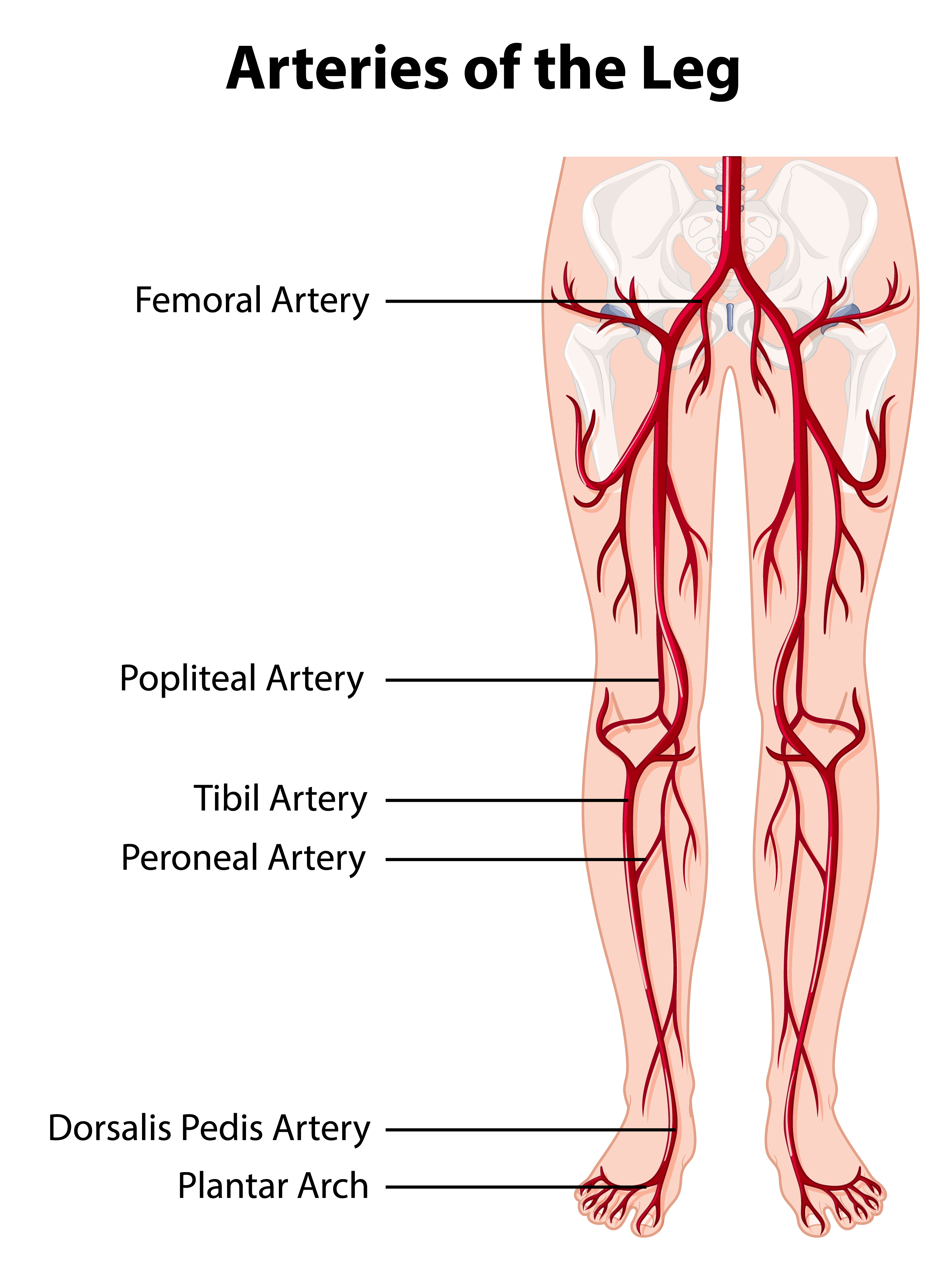
There is a point at which the anterior and posterior arterial circuits of the brain unite or anastomose. Fibular (peroneal) the fibular artery of the leg. Small branches dive into the heart muscle to. This is an online quiz called arteries of the body there is a printable worksheet available for download here so you can take the quiz with pen and paper. Constricted arteries oppose blood flow, and more pressure is required to push blood.
Arteries And Veins Of The Leg Educational Diagram Download Free Vectors Clipart Graphics Vector Art from static.vecteezy.com If your leg arteries are badly blocked, you may develop foot pain while resting or a sore that won't heal. After receiving blood directly from the left ventricle of the heart, the. The capillaries connect the two types of blood. The two exceptions are the pulmonary and the umbilical arteries, which carry deoxygenated blood to the organs that oxygenate it (lungs and placenta. It originates from the heart and branches out into smaller arteries which supply blood to the head region (brachiocephalic artery), the heart itself (coronary arteries), and the lower regions of the body. Coronary arteries supply oxygenated blood to the heart muscle, and cardiac veins drain away the blood once it has been deoxygenated. A wire is moved through an artery in the leg up to the carotid artery, and a small wire tube, or stent is expanded inside a narrowing of the carotid artery. Arteries are blood vessels that carry blood away from the heart.
There is a point at which the anterior and posterior arterial circuits of the brain unite or anastomose.
We hope this picture blood circulation principal veins and arteries diagram can help you study and research. For more anatomy content please follow us and visit our website: Start studying review of arteries. Systemic arteries deliver blood to the rest of the body. The vertebral arteries, and the internal carotid arteries. This area is known as the circle of willis. The capillaries connect the two types of blood. Lungs (pulmonary), and arteries, veins, coronary and portal vessels (systemic). Heart anatomy coronary arteries diagram. Blood is transported in arteries, veins and capillaries. This allows blood to flow around the blocked artery to another artery nearby or to the same artery past the blockage, protecting the heart tissue from injury. Blood carried by arteries is usually highly oxygenated, having just left the lungs on its way to the body's tissues. Artery, in human physiology, any of the vessels that, with one exception, carry oxygenated blood and nourishment from the heart to the tissues of the body.